
Oral Mucocele in Pediatric Patients: Clinical Features and Surgical Management
Oral mucoceles are common benign lesions of the salivary glands, frequently encountered in pediatric dentistry. This article explores the clinical characteristics of mucoceles in children, current diagnostic approaches, and the surgical procedures recommended for effective management.
 Introduction
Mucoceles are mucous-filled cystic lesions primarily resulting from trauma to minor salivary glands. They commonly appear in children and adolescents, particularly on the lower lip. While they are benign and painless, their recurrence and interference with oral functions can necessitate surgical intervention. Early identification and appropriate treatment are essential to prevent complications and ensure optimal oral health outcomes.
➤ Etiology
Oral mucoceles typically arise from:
° Extravasation: due to trauma or biting, leading to mucin leakage into surrounding tissues.
° Retention: due to ductal obstruction causing mucous accumulation.
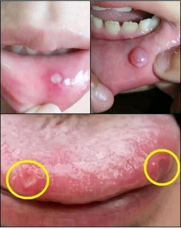
➤ Common Features in Pediatric Patients:
° Location: Predominantly on the lower lip, but may also appear on the buccal mucosa, ventral tongue, or floor of the mouth (ranula).
° Appearance: Bluish, translucent, and fluctuant swelling.
° Size: Ranges from a few millimeters to over 1 cm.
° Symptoms: Usually asymptomatic but may interfere with speech, chewing, or aesthetics.
Diagnosis
Diagnosis is primarily clinical, but additional tools may be required in atypical presentations:
° Clinical Examination: Inspection and palpation to assess size, consistency, and mobility.
° Ultrasound or MRI: For deeper lesions such as plunging ranulas.
° Histopathology: Confirms diagnosis post-excision.
Surgical Management
Surgical intervention is the preferred treatment for persistent or recurrent mucoceles.
➤ Common Techniques:
1. Conventional Excision
° Complete removal of the lesion along with associated salivary gland tissue.
° Local anesthesia is sufficient for most pediatric patients.
° Suturing may be required depending on the lesion’s size.
2. Marsupialization
° Typically used for large ranulas.
° Involves unroofing the lesion and suturing the edges of the mucosa to the surrounding tissue.
3. Laser Surgery
° CO₂ or diode lasers offer minimal bleeding and faster healing.
° Suitable for cooperative pediatric patients.
4. Micro-marsupialization
° A conservative technique for younger children with high recurrence rates.
➤ Postoperative Care
° Soft diet and good oral hygiene.
° Analgesics for discomfort.
° Follow-up to monitor for recurrence.
 Discussion
Oral mucoceles are frequently misdiagnosed or underestimated in pediatric populations. Due to their benign nature, some clinicians may prefer observation; however, surgical management offers definitive resolution and histopathological confirmation. Recurrence may occur if the associated glandular tissue is not entirely removed. Laser techniques show promise in reducing intraoperative bleeding and improving healing times, making them especially useful in pediatric dentistry.
 Conclusion
Oral mucoceles in pediatric patients, though benign, can impact oral function and quality of life. A comprehensive clinical evaluation followed by surgical excision remains the gold standard for treatment. Pediatric dentists must be familiar with both conventional and advanced surgical approaches to provide optimal care.
 References
 de Pontes, F. S., Neto, F. B., de Sousa, F. B., de Carvalho, M. G. F., & de Moraes Ramos-Perez, F. M. (2020). Clinical-pathological study of 206 cases of oral mucoceles in a Brazilian population. Medicina Oral, Patología Oral y Cirugía Bucal, 25(5), e566–e570. https://doi.org/10.4317/medoral.23368
Azenha, M. R., Bueno, R. B., & Silva, T. M. (2019). Management of oral mucoceles in pediatric patients: A review and case report. Journal of Clinical and Experimental Dentistry, 11(6), e571–e575. https://doi.org/10.4317/jced.55873
Baurmash, H. D. (2003). Mucoceles and ranulas. Journal of Oral and Maxillofacial Surgery, 61(3), 369–378. https://doi.org/10.1053/joms.2003.50071